Mouches Volantes
Floaters

Muscae Volitante
(Latin: "Flying Flies")

Floaters

Muscae Volitante
(Latin: "Flying Flies")

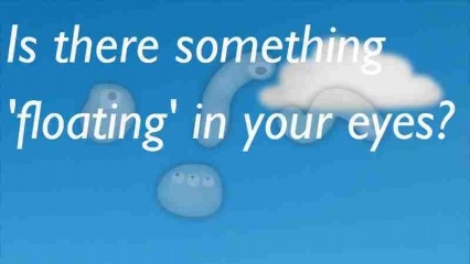
What are so called 'mouches-volantes' or flying flies?
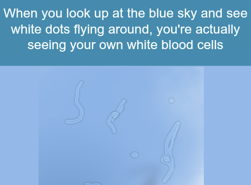
Are those a result of ever having watched into the sun?
Causes
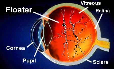
There are various causes for the appearance of floaters, of which the most common are described here. Simply stated, any damage to the eye that causes material to enter the vitreous humour can result in floaters.
Floaters can be a sign of retinal detachment or a retinal tear but in most cases (98% according to the National Health Service)[citation needed] it is simply age-related or due to natural change in the vitreous humour.
They are also called Muscae volitantes (Latin: "flying flies"), or mouches volantes
(from the French). Floaters are visible because of the shadows they
cast on the retina or refraction of the light that passes through them,
and can appear alone or together with several others in one's visual
field.
Floaters are deposits of various size, shape, consistency, refractive index, and motility within the eye's vitreous humour, which is normally transparent.
At a young age, the vitreous is transparent, but as one ages,
imperfections gradually develop. The common type of floater, which is
present in most persons' eyes, is due to degenerative changes of the
vitreous humour. The perception of floaters is known as myodesopsia, or less commonly as myodaeopsia, myiodeopsia, or myiodesopsia. They are also called Muscae volitantes (Latin: "flying flies"), or mouches volantes (from the French). Floaters are visible because of the shadows they cast on the retina or refraction of the light that passes through them, and can appear alone or together with several others in one's visual field. They may appear as spots, threads, or fragments of cobwebs, which float slowly before the observer's eyes. As these objects exist within the eye itself, they are not optical illusions but are entoptic phenomena. They are not to be confused with visual snow, although these two conditions may co-exist.
What are those floaty things in your eye?
Michael Mauser
Treatment
While surgeries do exist to correct for severe cases of floaters, there are currently no medications (including eye drops) that can correct for this vitreous deterioration. Floaters are often caused by the normal aging process and will usually disappear as the brain learns to ignore them. Looking up/down and left/right will cause the floaters to leave the direct field of vision as the vitreous humor swirls around due to the sudden movement. If floaters significantly increase in numbers and/or severely affect vision, then one of the below surgeries may be necessary.
Currently, insufficient evidence is available to compare the safety and efficacy of surgical vitrectomy with laser vitreolysis for the treatment of floaters. A 2017 Cochrane Review did not find any relevant studies that compared the two treatments.
Aggressive marketing campaigns are currently promoting the use of laser vitreolysis for the treatment of floaters.[18][19] No strong evidence currently exists for the treatment of floaters with laser vitreolysis. Currently, the strongest available evidence comparing these two treatment modalities are retrospective case series.
Surgery
Vitrectomy may be successful in treating more severe cases. The technique usually involves making three openings through the part of the sclera known as the pars plana. Of these small gauge instruments, one is an infusion port to resupply a saline solution and maintain the pressure of the eye, the second is a fiber optic light source, and the third is a vitrector. The vitrector has a reciprocating cutting tip attached to a suction device. This design reduces traction on the retina via the vitreous material. A variant sutureless, self-sealing technique is sometimes used.
Like most invasive surgical procedures, however, vitrectomy carries a risk of complications, including: retinal detachment, anterior vitreous detachment and macular edema – which can threaten vision or worsen existing floaters (in the case of retinal detachment).
While surgeries do exist to correct for severe cases of floaters, there are currently no medications (including eye drops) that can correct for this vitreous deterioration. Floaters are often caused by the normal aging process and will usually disappear as the brain learns to ignore them. Looking up/down and left/right will cause the floaters to leave the direct field of vision as the vitreous humor swirls around due to the sudden movement. If floaters significantly increase in numbers and/or severely affect vision, then one of the below surgeries may be necessary.
Currently, insufficient evidence is available to compare the safety and efficacy of surgical vitrectomy with laser vitreolysis for the treatment of floaters. A 2017 Cochrane Review did not find any relevant studies that compared the two treatments.
Aggressive marketing campaigns are currently promoting the use of laser vitreolysis for the treatment of floaters.[18][19] No strong evidence currently exists for the treatment of floaters with laser vitreolysis. Currently, the strongest available evidence comparing these two treatment modalities are retrospective case series.

Surgery
Vitrectomy may be successful in treating more severe cases. The technique usually involves making three openings through the part of the sclera known as the pars plana. Of these small gauge instruments, one is an infusion port to resupply a saline solution and maintain the pressure of the eye, the second is a fiber optic light source, and the third is a vitrector. The vitrector has a reciprocating cutting tip attached to a suction device. This design reduces traction on the retina via the vitreous material. A variant sutureless, self-sealing technique is sometimes used.
Like most invasive surgical procedures, however, vitrectomy carries a risk of complications, including: retinal detachment, anterior vitreous detachment and macular edema – which can threaten vision or worsen existing floaters (in the case of retinal detachment).


No comments:
Post a Comment